Skip to content

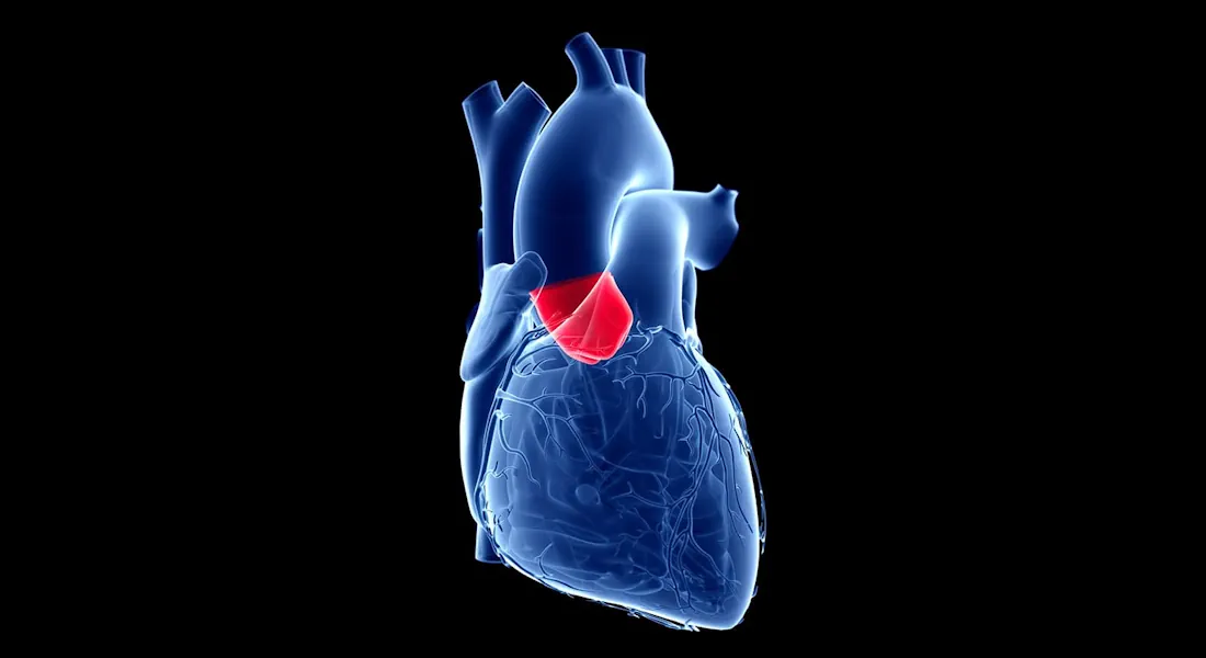
The aortic valve, located at the exit of the left heart chamber, plays a crucial role in ensuring blood flows efficiently from the heart into the aorta. Over time, this valve can become damaged, leading to aortic valve stenosis (blockage) or aortic valve regurgitation (leakage). When these conditions are advanced, valve replacement becomes necessary.

Approaches
Minimally Invasive

Mini (partial) sternotomy

Involves partially opening the chest bone.

Offers excellent access to the heart and great vessels.

Results in faster recovery and better cosmetic outcomes.

Mini-thoracotomy

A 4-6 cm incision between the 2nd and 3rd ribs without touching the chest bone.

Favoured by patients for its minimal invasiveness.

Suitable for patients with singular aortic valve problems, provided they meet specific anatomical criteria determined by a CT scan.

Totally endoscopic aortic valve replacement

Performed through a tiny incision (3-5 cm) without cutting or mobilising any bone.

Utilises an endoscopic camera for enhanced precision.

Open Heart

Full sternotomy

Involves a vertical incision along the chest to open the chest bone.

Provides the surgeon with a direct and unobstructed view of the heart and aortic valve.

Allows for comprehensive treatment of complex heart conditions that may not be suitable for minimally invasive techniques.


Conditions treated
Minimally Invasive
  • Aortic Valve Stenosis (Narrowing of The Aortic Valve)
  • Aortic Valve Regurgitation (Leaking of The Aortic Valve)
  • Combined Heart Conditions Requiring Surgical Intervention
Open Heart
  • Aortic Valve Stenosis (Narrowing of The Aortic Valve)
  • Aortic Valve Regurgitation (Leaking of The Aortic Valve)
  • Combined Heart Conditions Requiring Simultaneous Surgical Intervention (E.g. Heart Valve & Bypass Surgery At The Same Time)
  • Complex Anatomical Considerations That Preclude Minimally Invasive Options
What to expect
Before Surgery

Minimally Invasive & Open Heart

  • Thorough assessment including medical history, physical examination, and advanced imaging (e.g., echocardiogram, CT scan) to determine suitability for minimally invasive techniques.
  • Consultation with (Adj) Professor Theo Kofidis to discuss minimally invasive & open heart options, their potential benefits & risks, and if patient qualifies for minimally invasive surgery.
During Surgery

Minimally Invasive

  • General anaesthesia is administered.
  • Precise repair or replacement of the aortic valve through small incisions, minimising trauma to surrounding tissues.
  • Advanced technologies like Sutureless Aortic Valves (Perceval) and Cor-Knot Device (automated knot tying device) may be utilised for enhanced precision and efficiency.

Open Heart

  • General anaesthesia is administered.
  • Aortic valve repair or replacement performed through the open heart approach, involving opening the chest cavity.
  • Application of advanced surgical methods to ensure accurate procedure execution and optimal outcomes.
After Surgery

Minimally Invasive

  • Usually 1 day in ICU & 3-4 days in general ward.
  • Faster mobilisation and return to daily activities.
  • Follow-up appointments to monitor healing progress and overall cardiac health.

Open Heart

  • Usually 2 days in ICU & 5-7 days in general ward.
  • Slower mobilisation while in recovery.
  • Follow-up appointments to monitor healing progress and overall cardiac health.
Benefits
Minimally Invasive
  • Smaller incisions
  • Minimal scarring
  • Faster mobilisation & improved recovery
  • Less infection
  • Less blood loss
  • Less trauma
  • Less arrhythmia
  • Better lung function in the early stage
  • Better cosmetic effect
Open Heart
  • Suitability in certain complex & high-risk cases
Back To Top
No results found...